The human skeletal system comprises 206 bones, forming the body’s structural framework. It includes the axial and appendicular skeletons, providing support, protection, and facilitating movement while producing blood cells.
1.1 Overview of the 206 Bones in the Adult Human Body
The adult human body contains 206 bones, varying in size and shape. These bones form the structural framework, with 80 belonging to the axial skeleton and 126 to the appendicular skeleton. From the tiny ossicles in the ear to the large femur, each bone plays a unique role in supporting the body, enabling movement, and protecting internal organs.
1.2 Importance of the Skeletal System in the Body
The skeletal system is vital for providing structural support, facilitating movement, and protecting internal organs. It serves as an attachment point for muscles, enabling mobility, and houses bone marrow, which produces blood cells. Additionally, it stores minerals like calcium, maintaining the body’s overall health and stability. This system is essential for survival and bodily function.

Classification of Bones
Bones are classified into axial and appendicular categories. The axial skeleton includes skull, vertebral column, ribs, and sternum, while the appendicular comprises upper and lower limb bones.
2.1 Axial Skeleton (80 Bones)
The axial skeleton consists of 80 bones, including the skull, vertebral column, ribs, and sternum. It forms the body’s central framework, protecting vital organs like the brain and heart. The skull has 22 bones, the vertebral column 33, and the rib cage 24 ribs and the sternum, totaling 80 bones that provide structural support and muscle attachment points.
2.2 Appendicular Skeleton (126 Bones)
The appendicular skeleton comprises 126 bones, primarily in the upper and lower limbs, along with the shoulder and pelvic girdles. It facilitates a wide range of movements, providing attachment points for muscles and enabling effective locomotion and manipulation of objects. This skeletal section is crucial for mobility and structural support in the human body.

Major Bones of the Axial Skeleton
The axial skeleton includes the skull, vertebral column, rib cage, and sternum, totaling 80 bones. These bones provide structural support and protect vital organs, essential for overall body stability and function.
3.1 Skull Bones
The adult skull consists of 22 bones, including 8 cranial bones and 14 facial bones. These bones fuse during early adulthood, forming a protective structure for the brain and facial features. The cranium encases the brain, while facial bones support the jaw, cheekbones, and eye sockets, contributing to both structural integrity and sensory functions.
3.2 Vertebral Column
The vertebral column, or backbone, consists of 33 vertebrae in five regions: cervical, thoracic, lumbar, sacrum, and coccyx. The sacrum and coccyx are fused, resulting in 26 bones in adulthood. It provides structural support, protects the spinal cord, and allows flexibility for movement. Each region has unique features, with cervical vertebrae supporting the skull and lumbar vertebrae bearing significant body weight.
3.3 Rib Cage and Sternum
The rib cage and sternum are integral parts of the axial skeleton, comprising 12 pairs of ribs and the breastbone. The sternum connects the ribs, providing structural support and protection for vital organs like the heart and lungs. The rib cage facilitates breathing by expanding and contracting, with true ribs directly attaching to the sternum and false ribs connecting via cartilage. This structure is clearly illustrated in detailed PDF diagrams, showcasing the arrangement and connection points of the ribs to the sternum, aiding in anatomical study and identification;
Major Bones of the Appendicular Skeleton
The appendicular skeleton includes 126 bones, forming the upper and lower limbs. It comprises the humerus, radius, ulna, femur, patella, tibia, and fibula, as shown in detailed PDF diagrams;
4.1 Upper Limbs
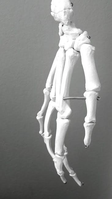
The upper limbs consist of 64 bones, including the humerus, radius, and ulna in the forearm, as well as carpals, metacarpals, and phalanges in the hands. These bones form the shoulder, elbow, wrist, and finger joints, enabling a wide range of movements. Detailed PDF diagrams illustrate their anatomical structure and connections, essential for understanding human anatomy.
4.2 Lower Limbs
The lower limbs consist of 64 bones, including the femur, patella, tibia, fibula, tarsals, metatarsals, and phalanges. These bones form the hip, knee, ankle, and toe joints, providing stability and enabling locomotion. PDF diagrams detail their structure, showcasing how they support the body’s weight and facilitate movement through a complex network of joints and bone connections.
Types of Bones Based on Shape
Bones are classified into five types: long, short, flat, irregular, and sesamoid. Long bones, like the femur, support body weight. Flat bones, such as skull bones, protect internal organs, while irregular bones, like vertebrae, have unique shapes for specific functions.
5.1 Long Bones
Long bones, like the femur and humerus, are cylindrical with a shaft and ends. They consist of compact bone for strength and spongy bone for shock absorption. These bones support body weight, facilitate movement, and are found in the limbs, playing a crucial role in mobility and structural support.
5.2 Short Bones
Short bones are cube-shaped and provide stability with limited movement. Found in the wrists, ankles, and feet, they absorb shock and distribute weight. Their dense structure, with a thin layer of compact bone and spongy bone inside, allows for durability while maintaining flexibility in joints, making them essential for balance and locomotion.
5.3 Flat Bones
Flat bones are thin, plate-like structures that protect internal organs and provide surfaces for muscle attachment. Found in the skull, ribs, sternum, and pelvis, they consist of two layers of compact bone enclosing spongy bone. Their broad, flat surfaces allow for efficient protection of vital organs, such as the brain and heart, while supporting overall body structure.
5.4 Irregular Bones
Irregular bones have unique, complex shapes that enable specialized functions. Examples include vertebrae, skull bones, and the pelvis. These bones protect internal organs, such as the brain and spinal cord, and provide attachment points for muscles and ligaments. Their intricate structures combine compact and spongy bone tissue, offering strength and flexibility where needed.

Bone Tissue and Structure
Bone tissue consists of compact bone, which is dense and homogeneous, and spongy bone, characterized by small, needle-like structures. Together, they provide strength, flexibility, and support to the body.
6.1 Compact Bone
Compact bone forms the dense, outer layer of bones, providing strength, protection, and support. It is composed of tightly packed collagen fibers and minerals like hydroxyapatite. This hard, smooth tissue is found in the shafts of long bones, such as the femur, and aids in movement by reducing friction between bone surfaces.
6.2 Spongy Bone
Spongy bone, also known as cancellous bone, is a lightweight, porous tissue found at the ends of long bones and within flat and irregular bones. It contains bone marrow, which produces blood cells, and its open structure allows for flexibility and adaptability while maintaining strength. This tissue is crucial for shock absorption and supporting the body’s movements.
Function of Bones in the Body
Bones serve multiple functions, including providing structural support, protecting vital organs, facilitating movement through joints, and producing blood cells in bone marrow.
Bones also store minerals like calcium and phosphorus, essential for overall health and bodily functions.
7.1 Support and Movement
Bones act as the body’s framework, enabling upright posture and movement through joints. They work with muscles to facilitate actions like walking, running, and sitting, while providing structural stability.
The skeletal system allows for a wide range of motion by connecting bones at movable joints, essential for daily activities and maintaining overall mobility.
7;2 Protection of Internal Organs
Bones safeguard vital organs, such as the brain, heart, and lungs. The skull encases the brain, while the rib cage protects the heart and lungs; Vertebrae shield the spinal cord, ensuring vital systems remain secure and functional.

Bone Development and Growth
Bone development begins with cartilage templates that gradually ossify. Growth continues through childhood and adolescence, with bones reaching full size and fusing by adulthood.
8.1 Bone Formation Process
Bone formation, or ossification, begins with cartilage templates in most bones. Osteoblasts secrete bone matrix, which calcifies to form compact bone. This process starts in the fetal stage, gradually replacing cartilage. Intramembranous ossification forms flat bones directly from mesenchyme, while endochondral ossification uses cartilage models for long bones. Both processes ensure proper shape and strength, contributing to the adult skeleton’s 206 bones.
8.2 Bone Fusion During Adulthood
Bone fusion occurs naturally as people age, reducing the total number of bones from childhood to the adult count of 206. This process enhances structural stability and support, particularly in weight-bearing areas like the pelvis and vertebral column. For example, the sacrum and coccyx fuse to form a single pelvic structure, while some skull bones unite to improve cranial stability.
Diagrams and Visual Representations
Diagrams of the 206 bones provide a detailed visual representation of the human skeletal system, categorizing bones into axial and appendicular groups for educational purposes.
9.1 PDF Diagram of the 206 Bones
A PDF diagram of the 206 bones offers a comprehensive visual guide, categorizing bones into axial and appendicular skeletons. It includes detailed labels and anatomical classifications, aiding students in identifying and studying each bone’s structure and location within the human body. These diagrams are often interactive, providing a deeper understanding of skeletal anatomy for educational purposes.
9.2 Labeling and Identification of Major Bones
PDF diagrams provide a detailed visual guide for labeling and identifying the 206 bones in the human body. These diagrams categorize bones into axial and appendicular skeletons, offering clear labels and classifications. They are essential tools for anatomy students, facilitating the study of complex skeletal structures and their functions through interactive and educational visual representations.
Educational Resources for Anatomy Students
PDF diagrams, printable bone charts, and interactive 3D models are essential tools for anatomy students, offering detailed visualizations and classifications of the 206 bones for comprehensive study.
10.1 Printable Bone Charts
Printable bone charts are valuable tools for anatomy students, offering detailed diagrams of the 206 bones in the human body. These charts provide clear visualizations of bone structures, aiding in identification and labeling. Available in PDF formats, they are ideal for study, classroom use, and quick reference, helping students grasp complex anatomical details effectively and efficiently.
10.2 Interactive 3D Models
Interactive 3D models provide a dynamic way to explore the human skeletal system, allowing users to rotate, zoom, and label bones virtually. These tools are particularly useful for anatomy students, offering a comprehensive view of bone structures and their relationships. Available online, they enhance learning by enabling hands-on interaction with detailed, accurate representations of the 206 bones in the body.
Common Misconceptions About Bones
A common myth is that the number of bones in the human body changes with age, but adults always have 206 bones, varying in size and shape.
11.1 Myth vs. Fact About Bone Count
A common myth is that the number of bones in the human body changes with age, but adults always have 206 bones. This misconception arises as some bones fuse during growth, such as the skull and vertebrae. However, the total count remains consistent, varying only in size and shape to perform different functions.
11.2 Bone Health and Maintenance
Bone health is crucial for overall well-being. A balanced diet rich in calcium and vitamin D, along with regular exercise, supports bone strength. Smoking and excessive alcohol can weaken bones, increasing fracture risks. Maintaining a healthy lifestyle helps prevent conditions like osteoporosis, ensuring long-term skeletal integrity and mobility.
The human skeletal system, comprising 206 bones, is vital for structural support, movement, and organ protection. Understanding its structure and maintenance is essential for overall health and longevity.
12.1 Summary of the 206 Bones
The adult human skeleton consists of 206 bones, varying in size and shape, from the tiny ossicles in the ear to the large femur. These bones form two main divisions: the axial skeleton (80 bones) and the appendicular skeleton (126 bones). Together, they provide structural support, protect vital organs, and enable movement, forming a complex framework essential for human function and survival.
12.2 The Role of Bones in Overall Body Function
Bones serve as the body’s structural foundation, enabling movement, protecting vital organs, and storing essential minerals like calcium and phosphorus. They also produce blood cells in the bone marrow, supporting the immune system. By providing attachment points for muscles, bones facilitate locomotion and maintain posture, playing a crucial role in overall bodily function and vitality.


